How AI Is Redefining Lung Cancer Screening Precision
Expert Perspectives · Waqas Haque, MD, MPH · 2025-12-15
In this edition of The Way Ahead: The Convergence of Technology and Cancer Care, Waqas Haque, MD, MPH, second-year Hematology/Oncology Fellow at the University of Chicago, explores how artificial intelligence (AI) is reshaping the precision and equity of lung cancer screening. Dr. Haque traces the field’s evolution, highlights emerging tools and real-world validation, and outlines how these advances are building a more accurate, accessible, and ethically grounded pathway from early detection to intervention.
Challenges and Gaps in Lung Cancer Screening
Lung cancer remains the leading cause of cancer death worldwide, but the landscape of early detection is undergoing a seismic shift. For years, screening guidelines relied heavily on traditional risk models that have left significant gaps, and the global incidence of lung cancer continues to rise due to the industrialization of societies.
As we near the end of 2025, a convergence of new research and deep learning validation is addressing these gaps. From the introduction of temporal tracking models to the validation of the “Sybil” tool in diverse populations, AI is showing its clinical necessity in cancer screening.
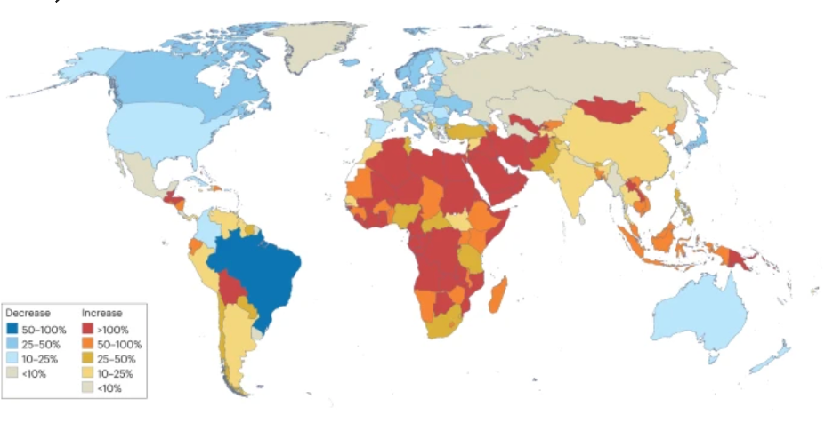
Changes in the number of individuals who smoke, by country (1990-2019), from Leiter et al (2023)
The Limit of Linear Regression
To understand where we are going with cancer screening, we have to understand where we have historically struggled. As highlighted in a recent Nature publication, traditional linear regression models often struggle to process and interpret the complex, high-dimensional data inherent in modern lung imaging. These older models frequently result in high false-positive rates (FPRs), causing unnecessary anxiety and procedures for patients, while simultaneously missing cancers in “lower-risk” populations. With the United States Preventive Services Task Force (USPSTF) guidelines evolving, there is a need to close the gap between cancers we screen for and the overall success rate of those screenings.
Introducing the CNN-GRU Model
One of the most exciting technical developments discussed this year is the integration of Convolutional Neural Networks (CNN) with Gated Recurrent Units (GRU). According to recent findings, this hybrid model is becoming a recommendation for accurate lung cancer detection because it mimics how a radiologist thinks, but with greater computational power:
- CNN (spatial): Extracts spatial data from the image, analyzing the texture and shape of lung tissue.
- GRU (temporal): This is where data is no longer static. The GRU captures temporal relationships between medical image sequences. By analyzing multiple CT scans over time, it monitors changes in lung tissue and tumor growth velocities that a single snapshot might miss.
This combination enhances the model’s capacity to identify subtle patterns and progressions, offering a dynamic view of patient health rather than a static one.
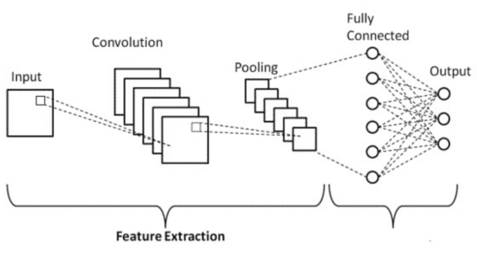
Convolutional neural network architecture, Jian et al (2025)
Sybil: Predicting Risk from a Single Scan
While CNN-GRU approach looks at changes over time, other breakthroughs are maximizing the utility of a single patient visit. We recently hosted Mary Pasquinelli, DNP, Nurse Practitioner and Director of the Lung Screening Program at the University of Illinois (UI) Health, on the Oncology Data Advisor podcast to discuss the significant research grants awarded to the University of Illinois (UI) Health. Her work focuses on “Sybil,” a deep learning model developed to predict future lung cancer risk from a single low-dose chest computed tomography CT (LDCT) scan.
Data presented at the American Thoracic Society (ATS) 2025 and recent reports regarding Sybil’s performance have shown remarkable potential. In a study evaluating over 21,000 individuals (aged 50-80) who underwent screening between 2009 and 2021, researchers followed outcomes through 2024. The results suggest Sybil can successfully bifurcate patients into two necessary groups:
- True low-risk individuals: Who may benefit from discontinuing further screening, reducing radiation exposure and healthcare costs.
- At-risk groups: Who should be encouraged to continue or accelerate screening.
Validating AI in Diverse Populations
Perhaps the most important update comes from the World Conference on Lung Cancer (WCLC) 2025. For AI to be ethical and effective, it must be trained on diverse datasets—new data has validated Sybil specifically within minoritized populations. In a study of 2,092 baseline LDCTs from UI Health’s lung screening program (2014-2024), the demographic breakdown was distinct:
- 62% non-Hispanic Black
- 13% Hispanic
- 4% Asian
Of these participants, 68 were diagnosed with lung cancer over follow-up periods ranging from zero to 10.2 years. The model’s ability to accurately predict risk in this predominantly non-Hispanic Black cohort is a step forward in health equity, ensuring that AI tools do not perpetuate the racial biases found in some historical clinical algorithms.

Dr. Waqas Haque (that’s me) speaks with Mary Pasquinelli, DNP, Nurse Practitioner and Director of the Lung Screening Program at UI Health
Other Exciting Developments in Lung Cancer Screening
- AstraZeneca’s CREATE study found its AI X-ray tool (qXR-LNMS) highly successful in lung cancer screening, with a 54.1% positive predictive value and 93.5% negative predictive value. The tool’s efficacy extended to non-traditional groups, like never-smokers, and is suggested for use before LDCT in resource-limited areas.
- AstraZeneca Korea deployed an AI-powered “Lung Health Check Bus” which screened 570 visitors, with 42% showing some form of lung abnormality. This real-world application demonstrates the utility of mobile AI screening in identifying lung issues in the general population.
- Researchers at UT Dallas developed an AI-powered breath biosensor that detects volatile organic compounds (VOCs) associated with thoracic cancers. In initial testing, the sensor correctly identified the cancer-linked VOCs in 90% of confirmed lung and esophageal cancer cases.

AstraZeneca Korea’s “Lung Health Check Bus” at the ABEXPO in Seoul!
The Path Forward
The transition from linear regression to deep learning and CNN-GRU architectures represents more than just a software update; it represents a fundamental change in how we view lung cancer pathology. By leveraging AI to see both the spatial details of a tumor and the temporal history of a patient, we are moving closer to a world where lung cancer is caught earlier, treated more effectively, and screened more equitably.
About Dr. Haque